Cold, Numb hands and regular neck pain began to be so intrusive that I was referred to specialist consultants to assess the conditions and what could be done to alleviate the them.
24 months since my hands began feeling numb and tingling. Not just an irritant, but something definitely amiss. Following GP’s appointments: hand specialist reviews: carpal tunnel elimination; Neurology assessment: traumatic MRI head and neck scans; posterior decompression (growth on the spine) diagnosis (Thank you QE, Dr Littleton and Seddigh): spinal surgery (thank you Mr Metcalfe), physiotherapy (Thank you Gina) and regular exercise, the symptoms are reducing. Shoulder pain is much less frequent, neck pains less painful, hands and fingers cold, but less frequently numb and fewer pins and needles.
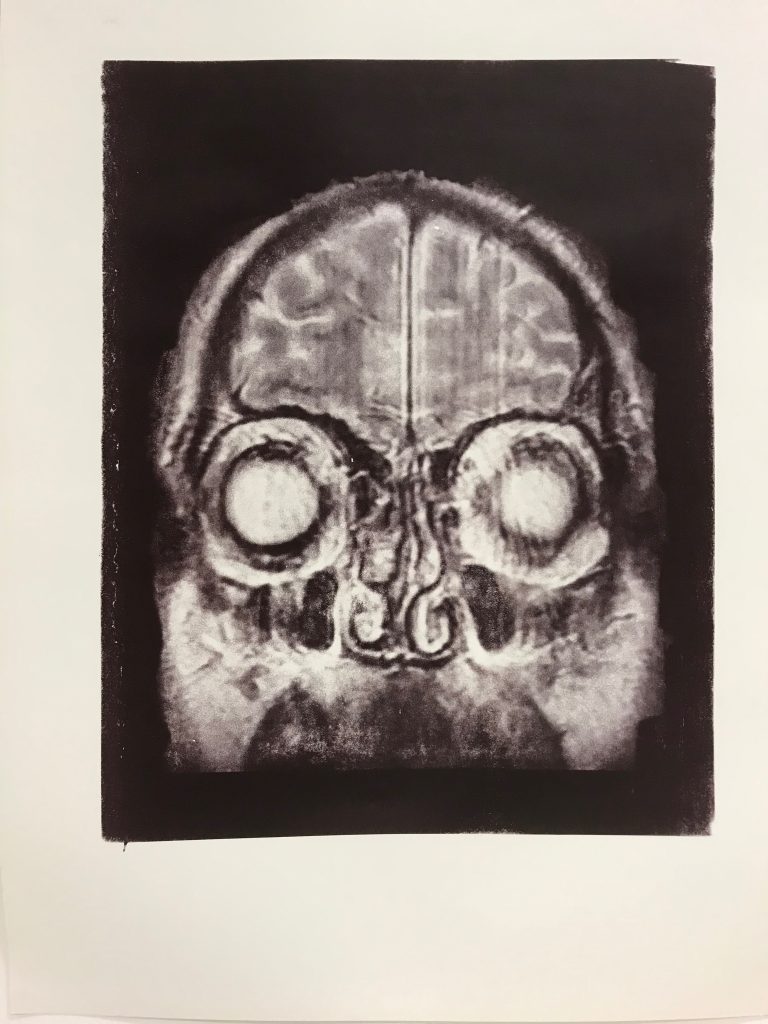
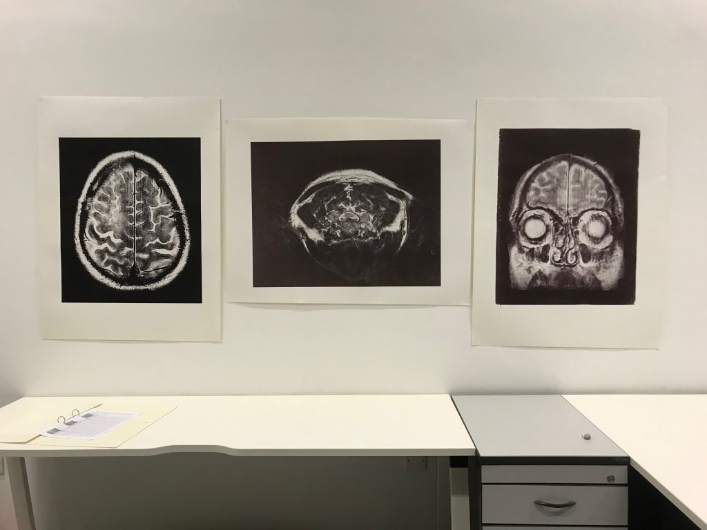
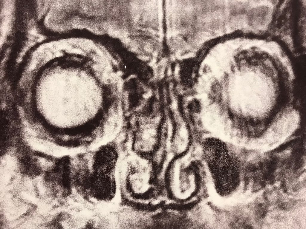
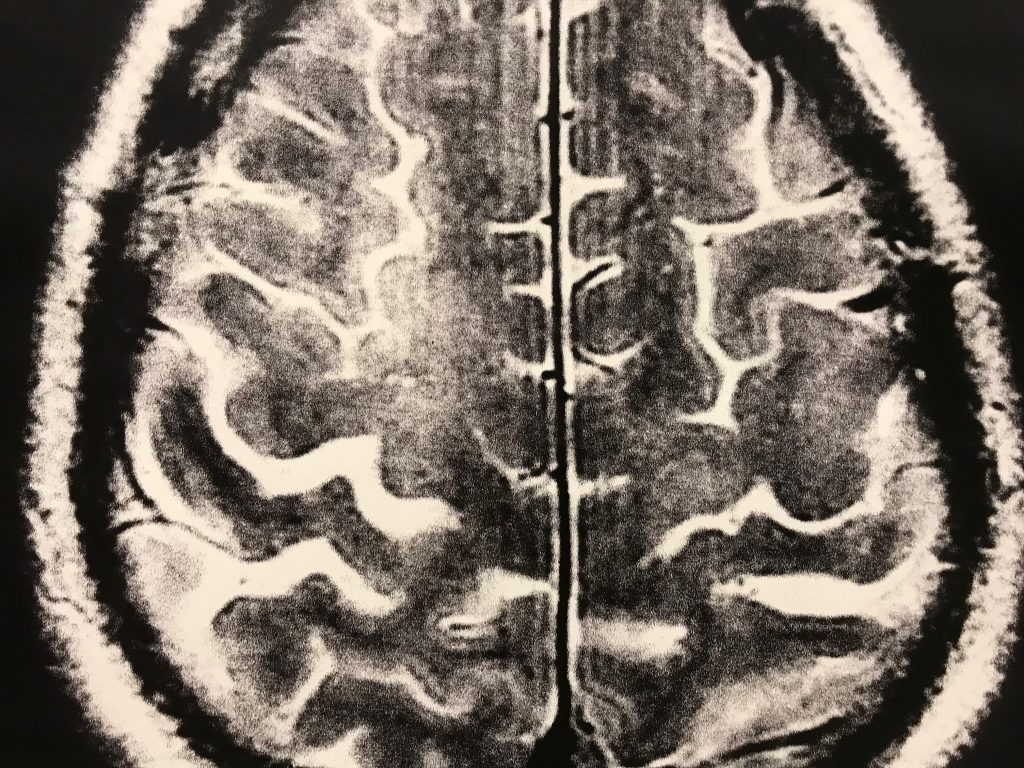
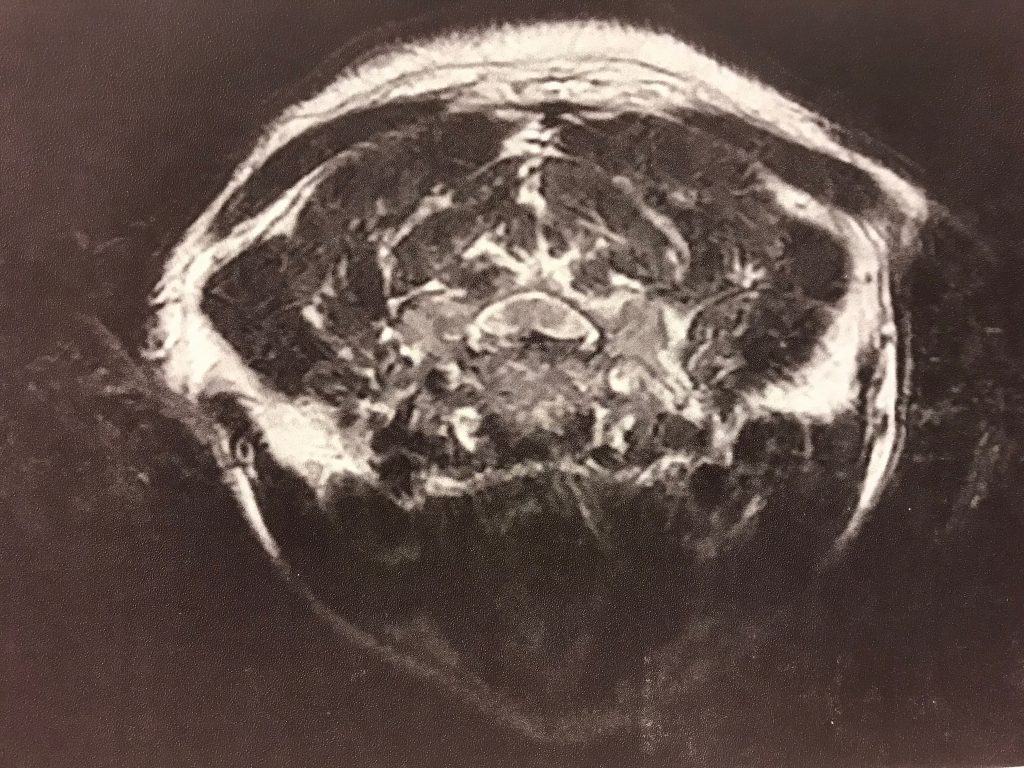
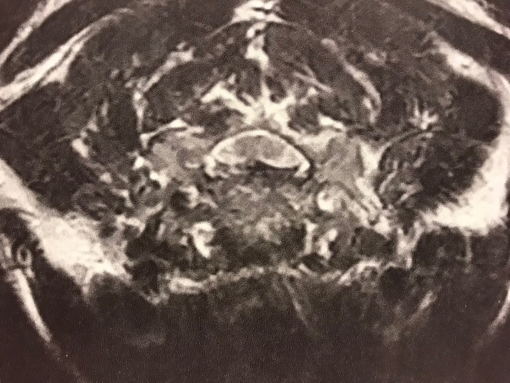
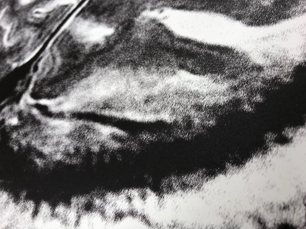
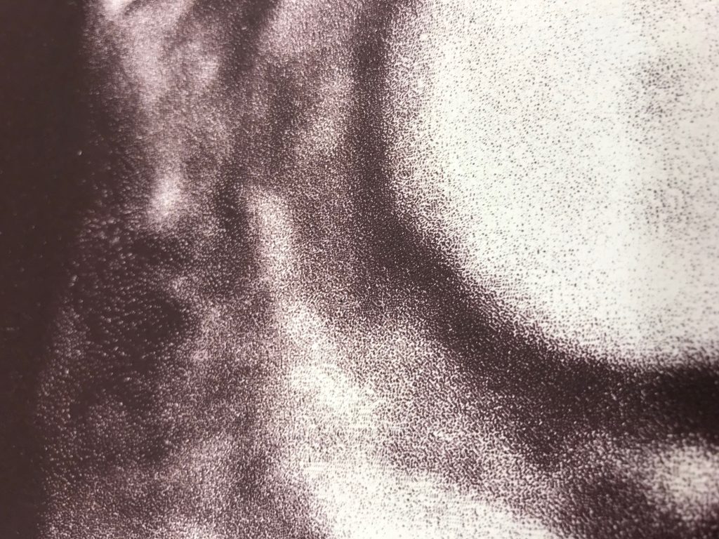
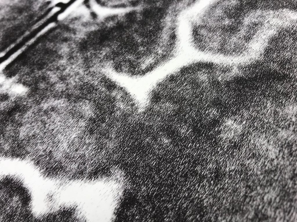
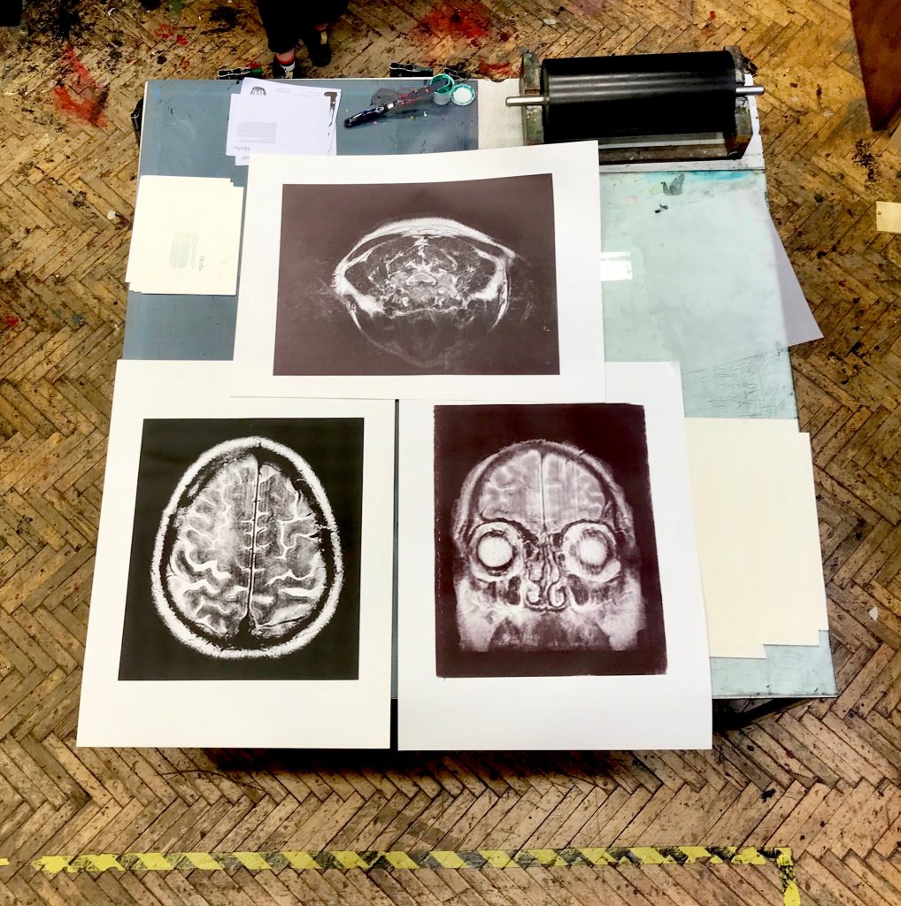
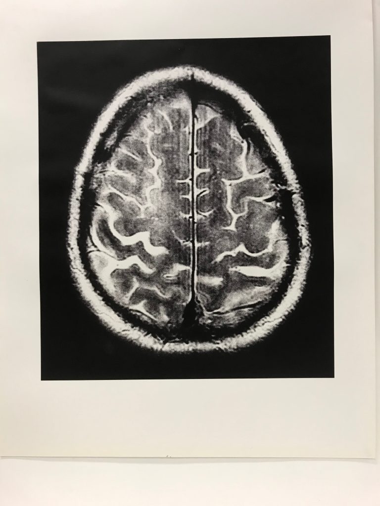
After the operation to remove growth at vertebrae’s C3 & 4, I accessed my pre-operation scan images. ‘Seeing inside myself’ for the first time inspired the making of a self portrait. Something I had never embarked upon before. Selecting ‘meaningful’ scans of neck, growth, head and brain I enlarged the small electronic images and applied a bitmap (black and white) matrix to give a texture and the ability to be silk screen printed at scale. The resultant three large scale printed images created an interior self portrait.
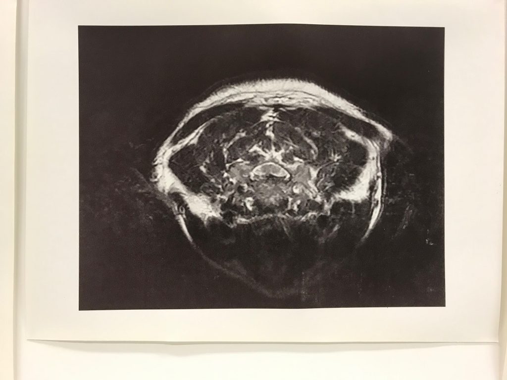
I had not shared the self portraits until, over an informal supper, I was introduced to an anatomy education expert. I could not resist enquiring about the diagnostic process and my anatomical make up. On showing my self portraits an enthusiastic informed discussion took off, where many of the questions I had been asking about the connectivity of my nervous system were clarified. Her enthusiasm for anatomy and ‘seeing into’ the neurorogical make up I had reflected, brought forth her knowledge of how brain and pain connect. Her ability to access online medical information through applying professional, rather than lay, language along with her engaging sharing of information made clear to me what vertebrae connected to which nerves. Which parts of the brain are responsible for which bodily function, and the relevant size of brain tissue for amount of sensory information.

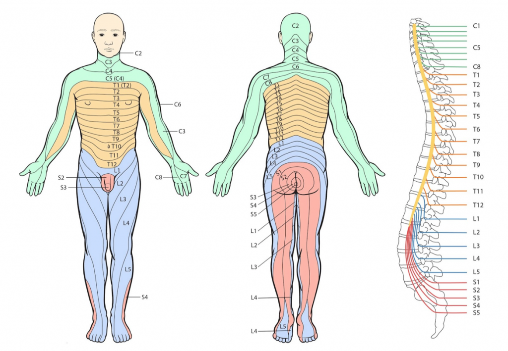
To explain how different parts of the brain process and control she brought up the ‘Cortical_homunculus’. This is a distorted representation of the human body, based on a neurological “map” of the areas and proportions of the human brain dedicated to processing motor, or sensory functions, for different parts of the body. Taking this further the nervous system is connected through dermatromes. Dermatrome, in anatomical terms, is an area of skin innervated by sensory fibers from a single spinal nerve.
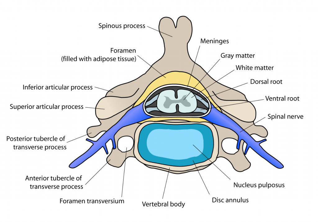
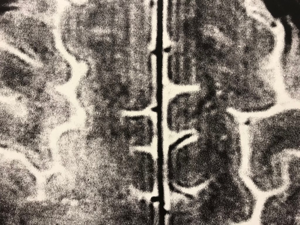
This healthy symmetrical cross section shows how the spinal chord is protected and the spinal nerves are cushioned. There are a number of online examples of MRI scans in the Radiopedia platform which aims to create the best radiology reference the world has ever seen and to make it available for free, for ever, for all.
This simple, but clearly explained description of the causes of nerve pains from the neck that had led to my posterior decompression surgery was immensely useful. My anatomical supper expert had been able to explain as she had time. She was not under medical service pressure to move onto the next patient.
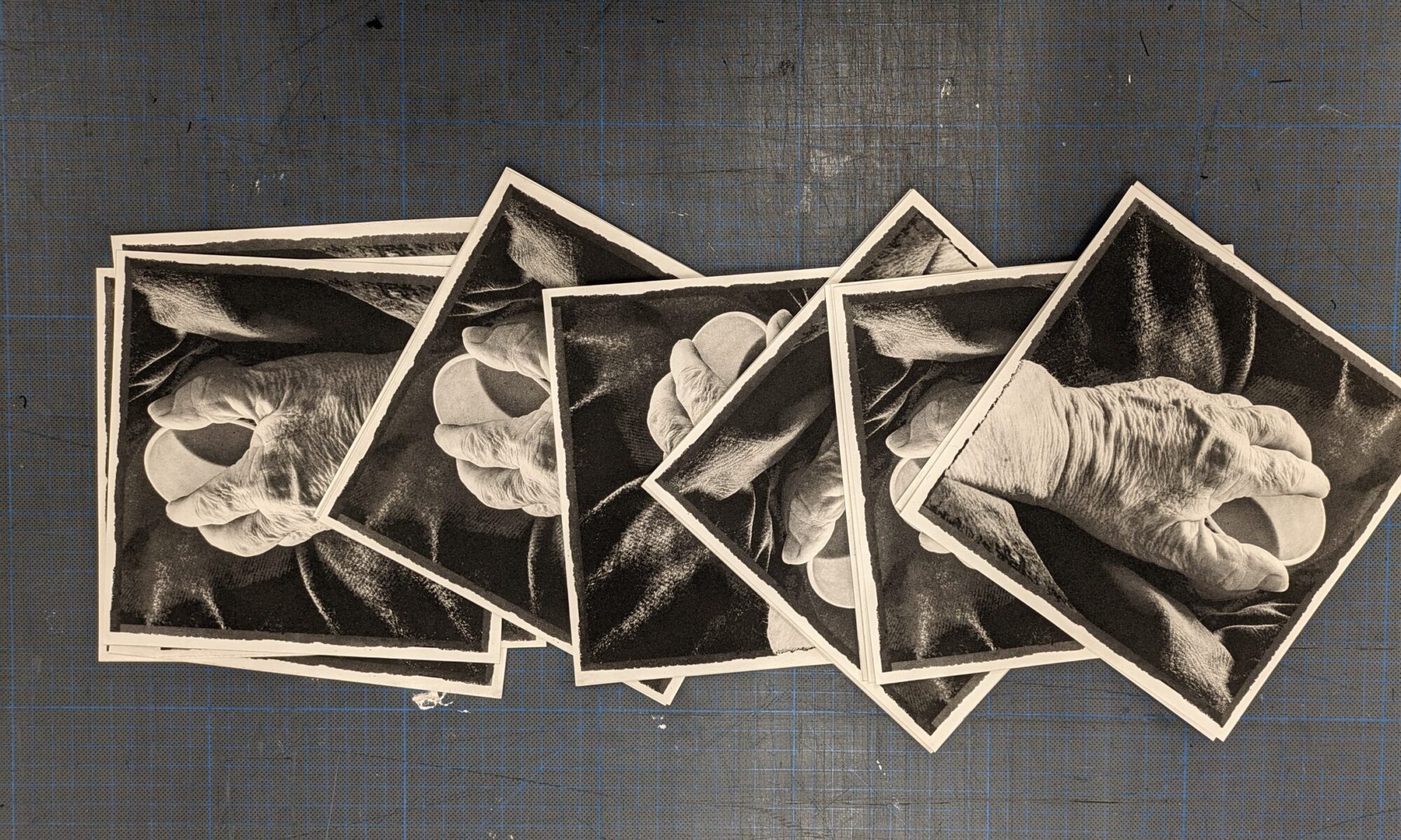
We returned to the printed self portraits and drew connection between the binary transmission of signals to, from and across the brain with the binary marks of information required to capture and print the interior self portrait images.
Our anatomy evening came to an end with the sharing of Leonardo Da Vinci’s 500th Anniversary catalogue, published this week and celebrated across the land and in the Birmingham Museum and Art Gallery. Da Vinci’s drawings laid the foundations for anatomical study and the knowledge base of today. Current Magnetic Resonance Imaging techniques allow detailed images of inside the live, human body to inform medical diagnosis and treatment. I am eternally thankful for the medics and my anatomical expert for enabling me to create my interior self portraits.